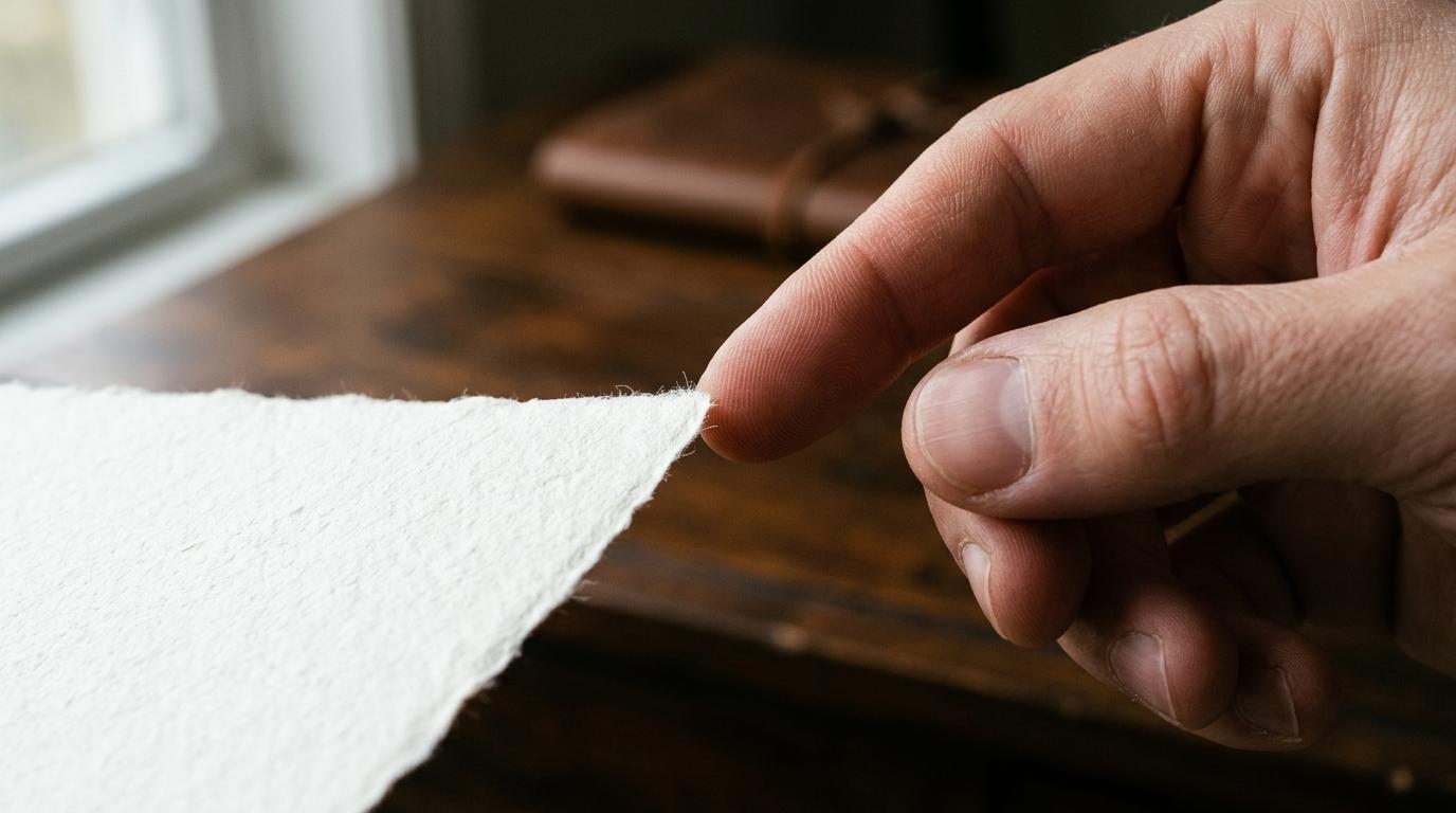
Few minor domestic injuries produce the immediate, breathless fury of a paper cut. Slicing through an envelope or turning a magazine page, a momentary slip leaves an almost invisible slit along the side of an index finger. There is rarely an outpouring of blood. There is no severed tendon, no deep muscular trauma, and no systemic emergency. Yet within seconds, a fierce, high-pitched, burning sting flares across the hand, followed by a persistent, throbbing ache that refuses to subside for hours or even days. Washing hands with warm water feels like acid, touching a keyboard sends sharp electric jolts through the palm, and even a mild breeze across the exposed skin triggers intense annoyance.
By contrast, slicing a finger while dicing vegetables with a sharp kitchen knife often feels duller and less lingering. While a knife cut bleeds profusely, demanding pressure and bandages, its acute stinging sensation frequently dissipates far faster than the persistent misery of a trivial paper slice. This discrepancy is neither psychological exaggeration nor an evolutionary fluke. It is the direct consequence of a perfect neurological and mechanical storm: extreme peripheral nerve density, microscopic material brutality, a failure of the physiological clotting cascade, and disproportionate cerebral amplification in the human sensory cortex.
The Paradox of the Trivial Wound: Why Paper Cuts Out-Sting Deep Lacerations
#To understand why a paper cut hurts so disproportionately, one must first dismantle the common assumption that wound size correlates linearly with perceived pain. In human trauma medicine, deep lacerations and blunt injuries certainly cause massive tissue destruction, but they stimulate different classes of sensory fibers and immediately activate systemic defense mechanisms that blunt localized burning.
A deep knife wound rapidly severs deep arterioles and venules, initiating an immediate deluge of whole blood. While alarming to behold, this hemorrhage serves critical evolutionary functions: it flushes foreign contaminants out of the wound bed, bathes damaged cells in natural analgesics and growth factors, and rapidly organizes into a platelet plug followed by a dense mesh of fibrin polymers. Within minutes, this fibrin clot hardens into a biological sealant, shielding vulnerable underlying nerve endings from the atmospheric environment.
A paper cut, conversely, is an anatomical anomaly: it is an exquisitely shallow wound. The razor-thin edge of a sheet of office paper rarely penetrates beyond the superficial stratum basale of the epidermis and the uppermost papillary dermis. Because the capillary loops in the papillary dermis are compressed and partially sheared rather than widely opened, the injury produces negligible bleeding. Without blood flow, there is no platelet aggregation, no fibrin polymerization, and no protective scab.
Instead, microscopic sensory nerve endings are left physically sheared open, raw, and completely unsealed. Every time air moves across the hand, microscopic moisture evaporates from the wound bed, concentrating local electrolytes and triggering osmotic shock. Every time the finger flexes, the severed dermal margins pull apart, mechanically restretching the injured nerve membranes. The paradox of the paper cut is that its superficial nature is precisely what makes it so agonizing: it destroys the cellular barrier while depriving the body of the biological seal needed to silence exposed pain receptors.
The Anatomical Landscape: Fingertip Nociceptor Density and Spatial Acuity
#The human hand is not merely a mechanical manipulator; it is the primary tactile sensory organ through which our species interrogates the physical universe. To allow humans to distinguish silk from wool, identify microscopic surface textures, and manipulate miniature tools with sub-millimeter precision, evolutionary selection packed the palmar surface of the hands—specifically the distal fingertips—with an extraordinary density of specialized sensory receptors.
Human skin is broadly divided into hairy skin and glabrous (hairless) skin. The glabrous skin of the fingertips contains four distinct classes of mechanoreceptors (Meissner corpuscles, Merkel cell-neurite complexes, Ruffini endings, and Pacinian corpuscles) intermingled with an extraordinarily dense lattice of free nerve endings known as nociceptors.
Nociceptors are high-threshold sensory neurons specialized in detecting noxious mechanical, thermal, or chemical stimuli capable of causing tissue damage. In the human back, thigh, or upper arm, nociceptor distribution is sparse, averaging fewer than 10 to 20 sensory terminals per square centimeter. On these broad surfaces, two sharp points placed two or three centimeters apart are frequently perceived as a single indistinct touch point—a clinical measure known as two-point discrimination.
On the human fingertip pulp, however, the anatomical architecture is entirely different:
- Innervation density exceeds 100 to 200 dedicated nociceptive nerve terminals per square centimeter.
- Two-point spatial discrimination on the distal finger pad is sharper than two millimeters, allowing the central nervous system to pinpoint the exact location of a microscopic cellular insult.
- Receptive fields of individual fingertip nerve fibers overlap extensively, meaning that even a sub-millimeter paper edge simultaneously cuts through dozens of distinct sensory units.
| Anatomical Body Region | Nociceptor and Tactile Density | Two-Point Spatial Discrimination | Dominant Sensory Role |
|---|---|---|---|
| Distal Fingertip Pad | 100 to 200 endings per sq cm | 1.5 to 2.5 mm | High-resolution tactile exploration |
| Perioral Region and Lips | 80 to 120 endings per sq cm | 2.0 to 3.0 mm | Ingestion, vocalization, thermal sensing |
| Palm of Hand | 40 to 60 endings per sq cm | 8.0 to 12.0 mm | Grasping, grip stabilization, load support |
| Dorsal Forearm | 15 to 25 endings per sq cm | 30.0 to 40.0 mm | Ambient environmental awareness |
| Mid-Back | 5 to 10 endings per sq cm | 40.0 to 70.0 mm | Coarse spatial orientation |
When a sheet of paper grazes an arm, thigh, or shoulder, it encounters a vast cellular plain with few sensory sentinels; the brain registers the brush as a trivial scratch. But when that exact same paper edge traverses the fingertip, it detonates a hyperdense minefield of specialized nociceptors, each sending emergency electrical volleys directly into the spinal cord.
The Cortical Homunculus: Why Your Brain Devotes Vast Neural Real Estate to Fingers
#The intensity of pain is never decided solely at the fingertip; it is processed, modulated, and interpreted within the primary somatosensory cortex of the brain, located in the postcentral gyrus of the parietal lobe.
In the 1930s, pioneering Canadian neurosurgeon Wilder Penfield mapped the functional organization of this cortical region by applying mild electrical stimulation to the exposed brains of awake patients undergoing epilepsy surgery. Penfield discovered that every region of the human body maps to a specific topographical band of cortical neurons, a neurological representation famously visualized as the sensory homunculus (Latin for little man).
The somatosensory homunculus is radically distorted. Rather than mirroring the physical proportions of the human skeleton, cortical surface area is distributed according to functional importance and receptor density. The trunk, legs, and back—despite accounting for the vast majority of physical body volume—occupy modest slivers of the cortical map. In striking contrast, the hands, thumbs, fingers, lips, and tongue occupy enormous, sprawling territories of the cerebral cortex.
This disproportionate representation, known as cortical magnification, means that millions of cortical neurons are continuously listening to sensory inputs arriving from your fingertips. When a paper cut occurs, the high-frequency action potentials generated by sheared digital nociceptors do not arrive at a neglected, low-priority processing hub. They flood into an expansive, highly sensitized neural computational matrix that scrutinizes every microvolt of change.
Understanding this cerebral real estate clarifies why the subjective emotional and cognitive burden of a paper cut feels overwhelming. Your brain is hardwired to care deeply about the integrity of its hands. From an evolutionary perspective, an injured or infected hand meant an inability to craft weapons, gather food, defend offspring, or ignite fire. The human central nervous system treats even a minor insult to the fingertip as a high-alert breach of the organism primary survival interface, illuminating why our understanding of human neurobiology and involuntary physiological reflexes consistently reveals evolutionary priorities embedded within sensory perception.
The Physics of the Paper Blade: Microscopic Serrations, Flexible Dullness, and Keratin Shear
#To understand the mechanical trauma of a paper cut, one must examine paper not as a flat, pristine plane, but through the lens of materials science and scanning electron microscopy. To human eyes, a fresh sheet of printer paper or envelope stock appears impeccably straight, smooth, and razor-sharp. Under 500x magnification, however, the edge of paper looks less like a surgical scalpel and more like a jagged, mangled saw blade constructed from compressed plant detritus.
Paper is manufactured by disintegrating wood into individual cellulose fibers, suspending them in water with chemical binders, bleaches, and mineral fillers (such as calcium carbonate and kaolin clay), and pressing the slurry through heated rollers. When an industrial blade slices these sheets to size, it does not create a molecularly smooth edge. Instead, it fractures, tears, and frays thousands of microscopic cellulose filaments, leaving an irregular, jagged, serrated boundary.
Furthermore, paper exhibits high mechanical flexibility and low structural rigidity compared to steel or glass:
- A steel scalpel or chef knife possesses high hardness and rigid mass. It enters the skin cleanly, parting cellular lipid bilayers and dividing keratin filaments with minimal lateral dragging.
- Paper, being flexible, buckles and bends as it strikes the stratum corneum (the tough, cornified outer layer of human skin).
- To penetrate the skin, paper requires lateral kinetic motion—a slicing drag across the surface.
- As the serrated, irregular paper edge drags across the epidermis, it does not part tissue cleanly. It grinds, tears, rips, and abrades cellular membranes, creating a ragged trench characterized by micro-avulsions, crushed keratinocytes, and frayed collagen bundles.
| Blade Material | Microscopic Edge Geometry | Penetration Mechanism | Primary Tissue Damage Profile |
|---|---|---|---|
| Surgical Steel Scalpel | Molecularly honed straight wedge | Direct perpendicular or linear parting | Clean linear division with minimal lateral cell lysis |
| Kitchen Utility Knife | Precision beveled steel edge | High-mass linear shear | Clean incised wound with prompt vascular transection |
| Standard 80 GSM Copy Paper | Ragged, torn cellulose fiber cluster | High-friction lateral sawing under light pressure | Microscopic tearing, ragged cell rupture, irregular trenching |
| Heavy Manila Cardstock | Coarse fibrous matrix with rigid binders | Blunt friction abrasion with localized crush | Broad epidermal avulsion, ragged micro-margins |
Because paper creates an irregular, jagged micro-wound rather than a clean cut, the total surface area of ruptured cellular membranes is significantly greater per millimeter of incision depth. Every crushed and torn cell spills its acidic intracellular contents into the extracellular interstitial space, creating a hostile biochemical bath that directly inflames adjacent nerve endings.
The Coagulation Deficit: Why Shallow Epidermal Wounds Stay Agonizingly Exposed
#Under normal physiological conditions, tissue trauma triggers the coagulation cascade—a complex biochemical symphony divided into primary and secondary hemostasis:
- Primary hemostasis: Vasoconstriction of severed blood vessels followed immediately by the adhesion, activation, and aggregation of blood platelets onto exposed subendothelial collagen.
- Secondary hemostasis: The sequential enzymatic activation of clotting factors (including thrombin and fibrinogen) along the intrinsic and extrinsic pathways, culminating in an insoluble, cross-linked mesh of fibrin threads that entraps red blood cells and solidifies into a stable clot.
This clot is not merely a biological cork to stop blood loss; it is a vital protective shield. As the clot hardens into a dry scab, it encapsulates the severed nerve endings, insulating them from ambient oxygen, atmospheric temperature fluctuations, and environmental contaminants.
In a typical paper cut, this protective mechanism completely fails because the incision terminates in the avascular epidermis or barely penetrates into the apex of the dermal papillae:
- The depth of the cut is insufficient to sever larger dermal blood vessels that carry adequate volumes of platelets and plasma clotting proteins.
- Only microscopic capillary loops are breached, which frequently collapse under the mechanical pressure of the paper stroke or release only a minuscule droplet of serous fluid.
- Without a robust volume of whole blood pooling within the wound channel, secondary hemostasis cannot proceed to full fibrin cross-linking.
Consequently, no protective scab forms over a paper cut. The severed ends of high-threshold nociceptive axons remain completely naked, exposed to the ambient atmosphere of the room. As ambient air passes over the unsealed wound, several destructive physiological phenomena occur:
- Evaporative cooling: Water continuously evaporates from the exposed interstitial fluid, dropping the local tissue temperature and triggering cold-sensitive transient receptor potential ion channels (such as TRPM8 and TRPA1) located on sensory axons.
- Hyperosmolar stress: As water evaporates, the concentration of extracellular sodium, potassium, and chloride ions rises steeply, destabilizing resting membrane potentials and causing spontaneous depolarization.
- Mechanical restretching: Because the fingertips are constantly in motion—grasping, typing, holding, and flexing—the ragged, unsealed epidermal edges continually slide past each other, repeatedly firing the exposed mechanoreceptors with every movement.
Two-Wave Pain Dynamics: Fast A-Delta Fibers Versus Lingering C Fibers
#Human peripheral pain transmission relies on two distinct classes of primary afferent nerve fibers, each with radically different structural properties, conduction velocities, and subjective sensations. A paper cut provides a textbook real-world demonstration of what neurophysiologists term dual-phase pain signaling.
Phase One: The Instantaneous Sting (A-Delta Fibers)
#The split second paper shears through the stratum corneum, the first wave of pain strikes. This sensation is sharp, bright, localized, and electric:
- Mediated by: A-delta fibers, which are lightly myelinated, medium-diameter axons (1 to 5 micrometers) that conduct action potentials at rapid velocities between 5 and 30 meters per second.
- Receptors activated: High-threshold mechanoreceptors and mechanothermal receptors (such as TRPV1 and Piezo2 ion channels) triggered by the violent physical tearing of cell membranes.
- Pathway: Signals travel rapidly up the peripheral nerves of the arm, enter the dorsal horn of the spinal cord (specifically laminae I and V), cross the midline, and ascend via the neospinothalamic tract directly to the ventral posterolateral nucleus of the thalamus, which relays the information straight to the primary somatosensory cortex.
- Evolutionary function: This rapid pathway informs the brain of the exact anatomical site of trauma within milliseconds, provoking an immediate, involuntary withdrawal reflex before the object can penetrate deeper into vital tendons or vasculature.
Phase Two: The Lingering, Throbbing Burn (C Fibers and Chemical Sensitization)
#Seconds after the initial sharp sting registers, a second, distinctly different type of pain begins to mount. This sensation is diffuse, dull, burning, aching, and deeply irritating:
- Mediated by: C fibers, which are small-diameter (0.2 to 1.5 micrometers), entirely unmyelinated axons that conduct action potentials at a sluggish pace between 0.5 and 2 meters per second.
- Pathway: C fibers terminate in laminae I and II (substantia gelatinosa) of the spinal cord dorsal horn and ascend primarily through the paleospinothalamic and spinoreticular tracts, projecting to the limbic system, insular cortex, and anterior cingulate cortex. This pathway governs the emotional suffering, distress, and unpleasantness associated with persistent pain.
The lingering burn of a paper cut is perpetuated by a vicious biochemical phenomenon known as peripheral sensitization, driven by the release of an inflammatory soup:
- Ruptured keratinocytes and damaged dermal cells spill intracellular adenosine triphosphate (ATP), potassium ions, and prostaglandins directly into the interstitial fluid.
- Local mast cells degranulate, releasing histamine and serotonin, which increase local capillary permeability and induce localized swelling.
- Axon reflexes cause unmyelinated C fibers to release neuropeptides, specifically Substance P and calcitonin gene-related peptide (CGRP).
- These chemicals bind to specific receptors on adjacent nerve terminals, dramatically lowering their activation threshold. A sensory receptor that previously required forceful mechanical deformation to fire is now so sensitized that even the microscopic friction of air molecules or a gentle thermal shift from room temperature triggers repeated, spontaneous action potentials.
Paper Chemistry vs Biology: Bleach, Sizing Agents, and Microscopic Debris
#For decades, popular folklore has asserted that paper cuts hurt because paper is impregnated with toxic chemicals that act as localized poisons. While modern commercial paper is not actively toxic in the sense of venom or systemic poisons, its biochemical composition does interact unfavorably with exposed human tissue.
Modern papermaking utilizes a vast array of chemical additives designed to enhance brightness, opacity, printability, and moisture resistance:
- Bleaching compounds: Wood pulp is routinely bleached using chlorine dioxide, hydrogen peroxide, or ozone to remove dark lignin residues and achieve high brightness. Trace chemical salts and oxidative residuals can remain embedded within the cellulose matrix.
- Sizing agents: To prevent liquid ink from feathering across the page, paper manufacturers coat fibers with sizing agents, typically alkyl ketene dimer (AKD), alkenyl succinic anhydride (ASA), or modified starches and rosin soaps.
- Mineral fillers: Up to 30% of standard copy paper by weight consists of fine particulate minerals, predominantly precipitated calcium carbonate, titanium dioxide, and kaolin clay, added to increase smoothness and opacity.
When the irregular edge of paper tears across the fingertip, it does not leave a pristine, sterile trench. Instead, it deposits microscopic debris—individual cellulose fibrils, mineral microcrystals, and trace sizing compounds—directly into the jagged epidermal wound bed.
Because the wound does not bleed sufficiently to wash out this microscopic detritus, these foreign particulates remain trapped against exposed nerve terminals. While they do not poison the bloodstream, they act as mechanical and osmotic irritants:
- Insoluble cellulose microfibrils physically prevent the jagged epidermal walls from collapsing back together and sealing.
- Particulate mineral fillers create localized hypertonic microenvironments that draw water out of damaged cells, exacerbating osmotic depolarization.
- Macrophages and neutrophils migrating to the injury site encounter foreign cellulose particles that cannot be rapidly broken down by human enzymes, amplifying localized release of inflammatory cytokines, tumor necrosis factor-alpha, and reactive oxygen species.
Evidence-Based First Aid: Liquid Bandages, Occlusion, and Rapid Receptor Pacification
#Understanding the exact neurobiology of a paper cut transforms first aid from a haphazard guessing game into a rational medical intervention. The primary objective of paper cut treatment is not to prevent catastrophic blood loss, but to mechanically replace the missing biological clot, eliminate evaporative cooling, and pacify sensitized nociceptors.
Step 1: Immediate Irrigation and Cleansing
#The instant a paper cut occurs, resist the instinct to suck on the finger. The human oral cavity harbors hundreds of distinct bacterial species, including Streptococcus and anaerobic pathogens, that can easily colonize an open epidermal wound.
Instead, wash the injured area under cool or lukewarm running tap water using a mild, non-drying soap. This mechanical irrigation flushes out trapped cellulose microfibrils, microscopic mineral fillers, and residual chemicals before they trigger deep macrophage activation. Pat the skin dry with a clean, lint-free cloth without aggressive rubbing.
Step 2: The Critical Principle of Artificial Occlusion
#Because the body cannot produce an immediate fibrin scab over a shallow paper cut, the definitive medical solution is to provide an immediate artificial barrier that mimics the biological seal of a blood clot:
- Topical Petroleum Jelly or Ointment: Applying a thin layer of pure white petrolatum creates an impermeable lipid barrier that halts evaporative water loss, prevents hyperosmolar electrolyte shifts, and keeps sensitized nerve endings in an isotonic environment.
- Medical Liquid Bandage (Cyanoacrylate Formulations): Liquid bandages or medical-grade cyanoacrylate skin adhesives represent the single most effective therapeutic tool for paper cut agony. When applied across the wound margins, the liquid polymerizes within seconds, chemically bonding the ragged epidermal edges together and creating an airtight, waterproof seal. By physically blocking air from contacting raw nerve terminals, liquid bandages immediately extinguish the burning sting.
Step 3: Mechanical Stabilization
#Even with a protective ointment or sealant, digital flexion continually pulls the wound edges apart whenever the hand moves. To prevent this mechanical disruption:
- Apply a snug, flexible adhesive bandage around the fingertip to restrict excessive dermal stretching.
- Avoid bulky dressings that trap excessive sweat, which can macerate the skin and delay re-epithelialization.
- For minor cuts, keeping the wound sealed under a clean bandage for 24 to 48 hours allows basal keratinocytes to migrate across the microscopic gap, re-establishing continuous epidermal integrity.
| Treatment Modality | Mechanism of Action | Time to Pain Relief | Impact on Healing Speed | Clinical Recommendation |
|---|---|---|---|---|
| Leaving Open to Air | None; permits continuous evaporation and desiccation | Slow (24 to 72 hours) | Delayed by crusting and ongoing cellular death | Not recommended |
| Sucking on Fingertip | Temporary saliva wetting; introduces oral bacteria | Transient (seconds only) | Increases risk of secondary bacterial paronychia | Strictly contraindicated |
| Petrolatum plus Bandage | Halts evaporation, maintains isotonic environment | Moderate (10 to 30 minutes) | Accelerates re-epithelialization by 40% | Highly recommended baseline |
| Liquid Skin Adhesive | Polymerizes into airtight synthetic scab across nerves | Immediate (under 60 seconds) | Optimal; locks wound edges in direct apposition | Gold standard for acute relief |
By combining prompt micro-irrigation with immediate polymer or petrolatum occlusion, you effectively short-circuit the neurological cascade that makes paper cuts so notoriously agonizing, silencing exposed nociceptors and allowing the body's remarkable cellular machinery to restore the skin barrier in peace.