It is a warm summer evening. You sit outdoors enjoying the breeze, completely unaware of the tiny insect landing softly on your forearm. Weighing barely two milligrams, the female mosquito steps across your skin with hair-thin legs, pierces your epidermis with a cluster of microscopic needles, drinks her fill of blood, and flies away undetected.
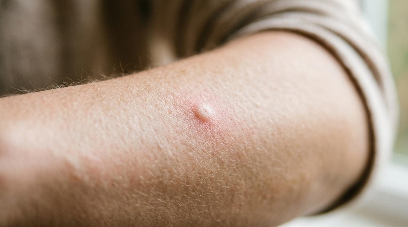
An hour later, the aftermath begins. First comes a faint tingling warmth, followed by a pale, swollen wheal surrounded by an expanding red halo. Then the itch arrives: an maddening, prickling, persistent sensory torment that demands immediate, aggressive scratching. You scratch the bump vigorously, feeling a brief, fleeting wave of relief—only for the itch to roar back twice as fierce moments later.
Why does a puncture wound smaller than a syringe needle cause such disproportionate cutaneous suffering? The answer lies not in the physical bite itself, but in the pharmacological cocktail left behind in your tissues. 
Mosquito bites itch because your immune system recognizes the foreign proteins in mosquito saliva and unleashes an acute allergic cascade driven by histamine, unmyelinated sensory nerve fibers, and a vicious neurobiological feedback loop in the spinal cord.
The Stealth Feeder: How a Mosquito Feeds
#Only female mosquitoes bite humans and other vertebrates. Male mosquitoes feed exclusively on plant nectar and sap. The female requires a dense concentration of dietary protein and iron to develop a clutch of eggs, making blood an absolute biological necessity for her reproduction.
When a mosquito lands on skin, she does not simply stab with a single blunt needle. Her mouthparts, known collectively as the proboscis, consist of a sophisticated fascicle of six specialized, interlocking stylets enclosed within a protective sheath called the labium:
Two serrated maxillae: These needle-thin blades feature microscopic teeth along their edges, which saw rapidly through the cornified outer layers of human skin with minimal tissue displacement.
Two mandibles: Sharp, stabilizing stylets that hold the puncture site open and guide the insertion.
The labrum: A hollow, groove-shaped channel through which blood is drawn up by muscular pharyngeal pumping.
The hypopharynx: A dedicated internal conduit through which the mosquito injects her saliva directly into the host's dermal tissue.
As the stylets penetrate the dermis, the mosquito probes the capillary bed, searching for a blood vessel in a process called solenophagy (direct vessel feeding). If she cannot immediately cannulate a vessel, she lacerates microscopic capillaries to create a tiny subterranean pool of blood, known as pool feeding or telmophagy.
The Salivary Weapon: Nature's Pharmacopeia
#Human blood is designed to clot almost instantly upon vessel injury. When a capillary wall is torn, platelets adhere to exposed collagen, activate, and aggregate to form a primary hemostatic plug. Simultaneously, the enzymatic coagulation cascade generates fibrin strands to cement the clot, while injured vascular endothelial cells release endothelin to constrict the vessel and limit blood loss.
If human blood clotted normally around the mosquito's proboscis, her microscopic feeding tube would clog within seconds, and she would starve.
To overcome this host defense, millions of years of co-evolution have transformed mosquito saliva into one of the most potent, complex anti-hemostatic cocktails in the biological world. A single female mosquito injects roughly one to five micrograms of saliva containing over a hundred distinct bioactive proteins, enzymes, and peptides:
Apyrase is a potent salivary enzyme that rapidly degrades adenosine diphosphate (ADP) and adenosine triphosphate (ATP) released by damaged cells. Because extracellular ADP is the primary chemical trigger for platelet activation and aggregation, apyrase effectively paralyzes the human body's first-line clotting response.
Factor Xa inhibitors and thrombin inhibitors directly block critical enzymatic checkpoints in the coagulation cascade, preventing prothrombin from converting into active thrombin and halting fibrin mesh synthesis.
Vasodilators, such as sialokinin (in Aedes mosquitoes) or tachykinin-like peptides, relax the smooth muscle lining of local arterioles and capillaries. By forcing the host's blood vessels to remain wide open, the mosquito dramatically increases blood flow to the puncture site, cutting her feeding time down to under four minutes.
Anesthetic peptides and anti-inflammatory modulators dampen immediate nociceptor firing, which is why you rarely feel the physical bite while the mosquito is actively feeding.
| Salivary Component | Primary Target | Biological Mechanism | Impact on Human Host |
|---|---|---|---|
| Apyrase | Platelet ADP Receptors | Hydrolyzes Free ADP into AMP | Prevents Platelet Aggregation |
| Sialokinin / Tachykinin | Arteriolar Smooth Muscle | Stimulates Nitric Oxide Pathway | Forces Capillary Vasodilation |
| Factor Xa Inhibitors | Coagulation Cascade | Blocks Prothrombinase Complex | Prevents Fibrin Clot Formation |
| Salivary Odorant Proteins | Immune Receptors | Binds Host Antigen Presenting Cells | Triggers IgE Allergic Sensitization |
The Immune Reaction: Mast Cells and Histamine Release
#Once the mosquito finishes her meal and flies away, the anti-hemostatic enzymes remain lodged in the extracellular matrix of your dermis. For the human immune system, these mosquito proteins are alien antigens.
In an adult who has been bitten previously, the immune system has already developed specific immunoglobulins—particularly Immunoglobulin E (IgE) and Immunoglobulin G (IgG)—tailored to recognize mosquito salivary antigens.
Resident in the dermis are mast cells: immune sentinel cells packed with granules containing pre-synthesized inflammatory mediators, foremost among them histamine. The outer surface of these mast cells is coated with high-affinity IgE receptors.
When the foreign mosquito salivary proteins bind and cross-link the specific IgE antibodies on the surface of adjacent dermal mast cells, an immediate signaling cascade is triggered. The mast cells undergo explosive degranulation, dumping high concentrations of histamine, leukotrienes, and prostaglandins into the surrounding interstitial fluid.
Histamine acts rapidly on two main targets:
Vascular endothelial cells: Histamine binds to H1 receptors on post-capillary venules, causing endothelial cells to contract and separate. This dramatically increases vascular permeability, allowing protein-rich fluid to leak out of the bloodstream into the surrounding dermal tissue. This localized fluid accumulation produces the raised, pale, edematous bump known as a wheal.
Local arterioles: Histamine stimulates the synthesis and release of endothelial nitric oxide, causing surrounding blood vessels to dilate. This rushes oxygenated blood to the area, creating the warm, erythematous red ring surrounding the bump, known as the flare.
Together, this response represents the classic Triple Response of Lewis: local redness, a surrounding flare, and an edematous wheal.
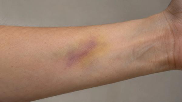
Understanding how our tissues manage acute microvascular trauma and cellular clearance is fundamental across dermatology. Readers exploring tissue resolution following capillary injury can examine how subcutaneous bruises change color over time, which details how macrophages systematically clear extravasated red blood cells and damaged hemoglobin in the dermis.
The Neurobiology of the Itch: From Skin to Spine to Brain
#While vascular swelling creates the physical bump, histamine's secondary target produces the sensory torment.
Embedded within the upper dermis, just beneath the epidermis, is a dense network of free sensory nerve endings belonging to unmyelinated C-fibers. A specialized subpopulation of these fibers, termed pruriceptors, is uniquely dedicated to detecting itch (pruritus).
Histamine released by degranulating mast cells binds directly to histamine H1 and H4 receptors located on these C-fiber terminals. Receptor binding activates an intracellular enzymatic pathway involving phospholipase C-beta-3 and transient receptor potential vanilloid 1 (TRPV1) channels, initiating an influx of sodium and calcium ions that depolarizes the sensory nerve.
Once depolarized, the nerve fires a barrage of electrical action potentials along peripheral sensory nerves to the dorsal root ganglion, entering the dorsal horn of the spinal cord.
Inside the spinal cord, pruriceptive signals synapse onto specialized second-order interneurons expressing gastrin-releasing peptide receptor (GRPR). These neurons project their axons across the midline and ascend the contralateral spinothalamic tract up to the thalamus, which routes the signals to the primary somatosensory cortex and the anterior cingulate cortex—the conscious brain centers responsible for the perception of itch and the emotional urge to scratch.
The Itch-Scratch Cycle: Why Scratching Makes It Worse
#When an itch strikes, your instinctive, immediate response is to drag your fingernails across the inflamed skin. For a split second, scratching feels profoundly pleasurable and relieving. But within moments, the itch returns with doubled ferocity. 
Why does scratching feel so good initially, and why does it inevitably backfire?
Scratching works initially by substituting pain for itch. Fingernails scraping across the epidermis create low-level mechanical pain and mild tissue damage. This pain signal travels up fast-conducting, myelinated A-delta nerve fibers to the spinal cord. 
Inside the dorsal horn, incoming pain signals stimulate specialized inhibitory interneurons (known as B5-I interneurons) that release glycine and GABA. These neurotransmitters act like a neural brake, temporarily inhibiting the GRPR itch-transmitting neurons. For a few seconds, the pain signal overrides and silences the itch signal, providing instant relief.
However, the brain responds to that sudden burst of pain by attempting to relieve it. Sensory centers in the brainstem, specifically the rostral ventromedial medulla, send descending pain-relief signals down the spinal cord by releasing the neurotransmitter serotonin (5-hydroxytryptamine).
While serotonin successfully blunts the sensation of pain, it has a disastrous side effect on itch:
Serotonin escaping into the spinal dorsal horn binds to 5-HT1A receptors located directly on the very same GRPR itch-transmitting neurons.
Instead of inhibiting them, serotonin activates these GRPR neurons, lowering their threshold and causing them to fire even more intensely in response to residual histamine.
Simultaneously, the physical trauma of fingernails clawing at the skin ruptures additional dermal mast cells and damaged epidermal keratinocytes, causing them to dump fresh waves of histamine, cytokines, and nerve growth factor into the bite site.
The result is a vicious neurobiological trap: itch leads to scratch, scratch triggers spinal serotonin and tissue damage, and serotonin amplifies the itch to an even higher level.
| Phase of Cycle | Primary Neurological Actor | Biochemical Event | Subjective Sensation |
|---|---|---|---|
| 1. The Trigger | Mast Cells and Dermal C-Fibers | Histamine Binds H1 Receptors | Intense Prickling Itch |
| 2. The Scratch | Fingernails on Epidermis | A-Delta Fibers Fire Pain Signals | Temporary Gate-Control Relief |
| 3. The Brain Response | Brainstem (Rostral Medulla) | Descending Serotonin Release | Pain Sensation Dampened |
| 4. The Amplification | GRPR Spinal Interneurons | Serotonin Binds 5-HT1A Receptors | Rebound Hyper-Sensitized Itch |
Why Bite Reactions Differ Between People
#Everyone knows individuals who seem to erupt into huge, golf-ball-sized welts from a single mosquito bite, while others notice barely a faint pinprick that vanishes in an hour. These vast disparities reflect differences in immunological sensitization, not bite intensity.
When humans are repeatedly exposed to mosquito bites over their lifetime, their immune system progresses through five distinct clinical stages:
Stage 1 (Non-sensitized): Observed in infants experiencing their first mosquito bites. Because the immune system has no immunological memory of salivary proteins, no antibodies exist. There is zero immediate or delayed reaction—no bump and no itch.
Stage 2 (Delayed reaction only): After repeated exposures, the immune system develops cell-mediated (type IV) hypersensitivity. Hours after the bite, a small, hard, itchy red papule appears, lasting several days.
Stage 3 (Immediate and delayed reaction): Common in older children and young adults. The body has now developed both IgE antibodies (causing an immediate wheal within 20 minutes) and cell-mediated immunity (producing a delayed, itchy nodule that lingers for days).
Stage 4 (Immediate reaction only): After hundreds of bites over years of adulthood, the delayed reaction begins to fade, leaving only a brief, transient immediate hive that disappears within a couple of hours.
Stage 5 (Natural desensitization): Following thousands of bites—frequently seen in indigenous populations or field biologists living in heavily infested regions—the body develops immunological tolerance (high IgG4 levels that outcompete IgE). Bites produce virtually no wheal, flare, or itch.
In rare individuals, an exaggerated immune hypersensitivity known as Skeeter syndrome produces extensive local cellulitis-like swelling, low-grade fever, and extreme pain, requiring medical evaluation and oral antihistamines.
Evidence-Based Relief: How to Stop the Itch
#Because the itch is driven by specific biochemical mechanisms, effective treatment relies on interrupting those pathways rather than yielding to the scratch reflex:
Immediate washing: Thoroughly wash the bite site with soap and cool water as soon as you notice it. This washes away any residual unabsorbed salivary proteins lingering on the skin surface.
Cold therapy: Applying an ice pack wrapped in a clean cloth directly to the bite for 10 to 15 minutes constricts local blood vessels, dramatically slowing the rate of fluid leakage and histamine distribution. Cold also activates TRPM8 cold-sensing receptors on sensory nerves, which directly inhibit pruriceptive transmission in the spinal cord without triggering the serotonin rebound cycle.
Topical hydrocortisone (1%): A mild corticosteroid cream applied to the bite suppresses local cytokine production, downregulates endothelial adhesion molecules, and stabilizes cell membranes, resolving both swelling and itch.
Oral second-generation antihistamines: For widespread or highly reactive bites, non-drowsy oral H1-antihistamines (such as cetirizine, fexofenadine, or loratadine) competitively block histamine from binding to peripheral C-fiber and endothelial H1 receptors.
Topical counter-irritants: Calamine lotion (zinc oxide) or low-concentration menthol creams soothe the skin by providing cooling sensory distraction without the tissue damage caused by fingernails.
Resisting the urge to scratch a mosquito bite is difficult, but understanding the neurochemical trap explains why discipline is essential. Every pass of a fingernail feeds the spinal serotonin loop, transforming a transient insect encounter into days of self-inflicted inflammation.