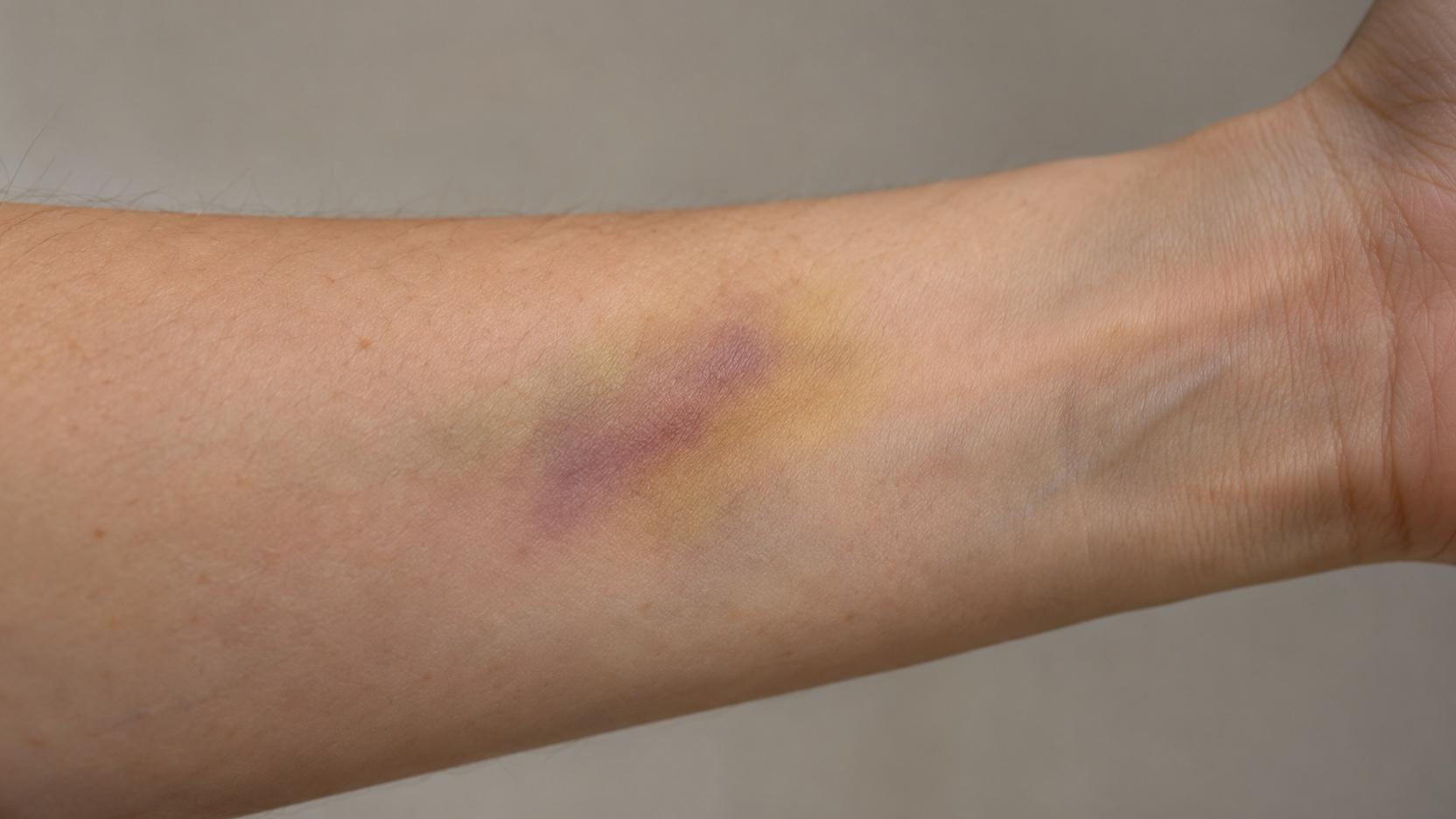
You bump your shin against a low coffee table or catch your bicep on a doorframe. At first, there is only a brief flash of pain, accompanied by a pale pink or faint red flush across the skin. By the next morning, that flush has darkened into an alarming, tender blotch of deep midnight purple and inky navy blue. Several days later, as the soreness begins to ease, the mark undergoes a bizarre, sickly transformation, turning a murky olive green. By the second week, it shifts into a vivid, glowing mustard yellow, then fades into a faint, rusty golden-brown shadow before finally vanishing completely.
This vivid chromatic journey is something virtually every human being has observed on their own body. Known in clinical medicine as an ecchymosis or contusion, a bruise is often described colloquially as bad blood pooling beneath the surface. Yet to dismiss a bruise as stagnant blood is to miss one of the most sophisticated, beautifully choreographed biochemical recycling operations in the human body.
The shifting colors of a bruise are not random marks of tissue decay, nor do they signal internal infection. They are the visible, real-time footprints of molecular enzymatic chemistry taking place in the microscopic spaces between your cells. As specialized immune cells called macrophages arrive at the injury site to clean up cellular wreckage, they systematically dismantle hemoglobin molecules through a precise series of metabolic reactions. Each stage of this breakdown yields a chemically distinct pigment molecule that absorbs and reflects specific wavelengths of light through the overlying translucent layers of human skin.
Understanding the biochemistry of a bruise transforms a mundane domestic injury into an extraordinary lesson in hematology, cellular recycling, and clinical pathology.
The Subcutaneous Leak: How Blunt Mechanical Force Creates a Contusion
#To understand why a bruise changes color, one must first examine what happens inside the tissue during the moment of mechanical impact. A bruise is fundamentally an internal vascular injury:
- Blunt Compressive Shear: Unlike a knife cut or an abrasion, which breaches the outer stratum corneum of the epidermis, a contusion is caused by blunt mechanical kinetic energy. When a hard object strikes the body, the kinetic shockwave compresses the skin and underlying subcutaneous fat against rigid bone or dense muscle fascia.
- Microvascular Rupture: While the elastic, flexible outer epidermis stretches and remains physically intact, the delicate, fragile microvasculature lying within the dermis and subcutaneous adipose tissue—predominantly capillary loops, arterioles, and postcapillary venules—cannot absorb the sudden shear stress. Their thin endothelial walls tear open.
- Extravasation of Whole Blood: Blood immediately escapes from the ruptured vessels into the surrounding interstitial fluid matrix, a process known as extravasation. Red blood cells (erythrocytes), blood platelets, and plasma fluids flood the spaces between collagen fibers and fat cells.
- Primary Hemostasis: Within seconds, exposed subendothelial collagen triggers platelet adhesion and the coagulation cascade, forming internal platelet-fibrin plugs that seal the damaged capillary walls and halt ongoing hemorrhage.
| Structural Layer | Anatomical Composition | Impact Response to Blunt Force | Role in Bruise Appearance |
|---|---|---|---|
| Epidermis | Cornified stratified squamous epithelium | Stretches elastically without tearing; remains sealed | Translucent optical window through which pigments are viewed |
| Papillary Dermis | Loose collagenous network with capillary loops | Superficial capillaries rupture; minor serous leakage | Early superficial pink and bright red hue |
| Reticular Dermis | Dense irregular collagen bundles and venules | Moderate vascular rupture; localized fluid edema | Traps pooled erythrocytes, establishing deep boundary |
| Subcutaneous Hypodermis | Adipose lobules, larger arterioles, and fascial planes | Large vessels shear; substantial blood extravasation | Primary reservoir for large hematoma pooling and color shifts |
Once the bleeding stops, the body faces an urgent biological crisis. Erythrocytes are designed to circulate continuously within a sealed, high-flow vascular network, where they deliver oxygen and clear carbon dioxide. When millions of red blood cells are trapped outside blood vessels in stagnant interstitial tissue, they are deprived of nutrients, oxygen, and metabolic clearance. Within hours, they begin to die, their cellular membranes degrade, and their internal cargo—billions of iron-rich hemoglobin molecules—spills into the extracellular space.
Day 0 to 2: Fresh Red to Deep Blue (Extravasation and Hemoglobin Deoxygenation)
#The first phase of a bruise is defined by the life cycle and oxygen saturation of hemoglobin. Hemoglobin is a complex, four-subunit globular protein that carries oxygen throughout the circulatory system. At the core of each subunit sits a specialized chemical coordination complex: a planar porphyrin ring known as heme, which cradles an atom of iron in its ferrous (Fe2+) state.
Hours 0 to 24: The Bright Red Flush
#In the immediate aftermath of blunt trauma, a fresh bruise typically appears bright red, reddish-pink, or tender crimson:
- Presence of Oxyhemoglobin: The red blood cells escaping from sheared dermal arterioles are packed with oxyhemoglobin—hemoglobin that is fully loaded with oxygen molecules bound to its iron atoms.
- Optical Properties: Oxyhemoglobin possesses a distinct molecular conformation that absorbs short blue and green wavelengths of light while reflecting long red wavelengths back through the epidermis, producing a vivid red appearance.
- Localized Hyperemia: In response to mechanical cell injury, local mast cells release histamine, causing acute vasodilation and increased blood flow to surrounding uninjured capillaries, adding to the red, flushed, slightly swollen perimeter.
Hours 24 to 48: The Deep Purple and Inky Blue Shift
#Within 24 to 48 hours, the bright red hue darkens dramatically into deep purple, dark navy, or blackish-blue:
- Rapid Oxygen Depletion: Because the pooled red blood cells are completely cut off from the respiratory circulation, their cellular metabolism rapidly exhausts all remaining oxygen molecules bound to the heme iron.
- Formation of Deoxyhemoglobin: As oxygen dissociates, oxyhemoglobin transforms into deoxyhemoglobin. This conformational change alters the electronic orbital configuration of the heme iron, causing the molecule to absorb red light far more strongly while reflecting darker blue and violet wavelengths.
- Light Scattering in Tissue: This dark blue appearance is amplified by a physical optical effect known as Rayleigh-Gans scattering. As ambient light penetrates through the translucent epidermis and superficial dermis, shorter blue wavelengths scatter more easily back toward the viewer eyes, while longer red wavelengths penetrate deeper and are absorbed by the dense, stagnant pool of trapped blood.
Day 3 to 6: Muddy Green (Heme Oxygenase and Biliverdin Cleavage)
#By the third or fourth day, the acute tenderness of the bruise begins to subside, and the dark purple center starts to undergo its most striking color transition: it morphs into a distinct, murky olive green.
This green transformation marks the arrival of the immune system cleanup crew. Tissue trauma triggers the release of chemical signaling beacons called chemokines, which summon large numbers of circulating monocytes from the bloodstream. Upon entering the bruised tissue, these monocytes mature into activated tissue-resident macrophages:
- Phagocytosis of Red Blood Cells: Macrophages engulf the fragile, dying red blood cells and free hemoglobin molecules via endocytosis, pulling them into specialized intracellular digestive chambers termed lysosomes.
- Heme Oxygenase-1 Activation: Inside the macrophage, an essential enzyme named heme oxygenase-1 (HO-1) goes to work on the released heme rings.
- Enzymatic Ring Cleavage: Heme oxygenase-1 breaks open the rigid, cyclic tetrapyrrole ring of heme at a specific alpha-methene bridge. This enzymatic cleavage converts the dark, cyclical iron-porphyrin structure into a linear, open-chain tetrapyrrole molecule called biliverdin.
- The Green Pigment: Biliverdin (derived from the Latin word bilis, meaning bile, and the French vert, meaning green) is a vividly green pigment. As macrophages process millions of heme molecules simultaneously, biliverdin accumulates within the extracellular interstitial fluid, imparting an unmistakably green hue to the healing tissue.
- Carbon Monoxide and Iron Release: During this cleavage reaction, the enzyme simultaneously ejects the central iron atom (releasing it for cellular storage) and generates a minute, physiological puff of carbon monoxide (CO), which acts as a localized anti-inflammatory gas signaling molecule.
| Chronological Stage | Typical Timeframe | Dominant Pigment Molecule | Chemical Precursor | Visual Color Observed |
|---|---|---|---|---|
| Acute Extravasation | Hours 0 to 24 | Oxyhemoglobin | Intact arterial erythrocyte cargo | Bright red or crimson flush |
| Deoxygenation Phase | Day 1 to 2 | Deoxyhemoglobin | Extravasated erythrocytes losing oxygen | Deep purple, dark blue, or blackish-navy |
| Porphyrin Cleavage | Day 3 to 6 | Biliverdin | Heme cleaved by heme oxygenase-1 | Olive green or murky sea-green |
| Biliverdin Reduction | Day 7 to 10 | Bilirubin | Biliverdin reduced by biliverdin reductase | Bright yellow, golden, or mustard hue |
| Iron Sequestration | Day 10 to 14+ | Hemosiderin | Ferritin aggregates and oxidized iron complexes | Faint golden-brown or rusty tan |
Day 7 to 10: Bright Golden-Yellow (Biliverdin Reductase and Bilirubin Generation)
#Around the end of the first week, the murky green coloration begins to fade, replaced by a radiant golden-yellow or warm amber hue that often forms an expanding ring around the outer borders of the contusion.
This yellow phase is driven by the immediate second step in the catabolic pathway:
- Biliverdin Reductase: Inside the macrophage cytoplasm, an enzyme called biliverdin reductase acts rapidly upon the newly formed green biliverdin molecules.
- Reduction into Bilirubin: Biliverdin reductase reduces the central carbon-carbon double bond of biliverdin, converting the green pigment into a yellow-orange tetrapyrrole pigment named bilirubin.
- The Molecule of Jaundice and Bile: Bilirubin is the exact same chemical pigment responsible for the classic yellow coloration of clinical jaundice (when liver clearance fails) and the golden-yellow coloration of normal bile.
- Optical Expression: Bilirubin strongly absorbs blue and violet light, reflecting bright, warm yellow wavelengths back through the skin. Because bilirubin is lipid-soluble, it diffuses easily through the extracellular fluid matrix, often spreading outward into a wide, diffuse yellow halo that surrounds the resolving core of the original bruise.
Day 10 to 14+: Brownish-Rust to Full Clearance (Hemosiderin and Macrophage Clearance)
#During the final phase of bruise resolution—typically occurring between day ten and day fourteen or beyond—the bright yellow tones slowly dull into a faint, rusty, golden-brown shadow.
This final chromatic stage is governed by the fate of the iron atoms that were stripped away from hemoglobin during the initial biliverdin cleavage:
- The Dangers of Free Iron: Free iron (Fe2+) is highly cytotoxic. If left unbound in tissues, free iron participates in the Fenton reaction, generating highly destructive hydroxyl free radicals that damage cell membranes and DNA.
- Ferritin and Hemosiderin Sequestration: To prevent oxidative tissue injury, macrophages immediately bind the liberated iron atoms to a protective spherical storage protein called apoferritin, forming ferritin. When massive quantities of iron are released at once—as occurs during a substantial contusion—macrophages package the excess iron into dense, insoluble crystalline complexes called hemosiderin.
- The Golden-Brown Stain: Hemosiderin is an iron-oxide-protein composite that exhibits an intrinsically golden-brown, rusty color. As macrophages gradually clear the water-soluble bilirubin through the lymphatic system, the insoluble, brown hemosiderin granules remain behind in the tissue for several extra days.
- Final Lymphatic Evacuation: Slowly, scavenger macrophages digest the remaining hemosiderin, export the iron into the bloodstream bound to transferrin (to be transported to the bone marrow for the synthesis of brand-new red blood cells), and migrate into regional lymph nodes. Once the last hemosiderin granules are cleared, the skin returns to its pristine, unblemished baseline.
Understanding this delicate microvascular and enzymatic dance highlights how our broader understanding of cutaneous microvascular fragility and coagulation cascades consistently shows that the human body treats even minor dermal disruptions with immense metabolic sophistication.
Accelerating Recovery: Ice, Compression, Elevation, and Evidence-Based Care
#While the biochemical cascade of a bruise is an automated internal process, clinical sports medicine and dermatological research have identified evidence-based protocols that can significantly minimize the initial size of a bruise and accelerate its clearance.
The standard first-aid framework is built around the fundamental laws of microvascular hemodynamics:
Phase 1: The First 48 Hours (Cold and Vasoconstriction)
#The single most critical window for managing a contusion is the first two days after impact:
- Immediate Cold Therapy: Apply an ice pack wrapped in a thin towel directly over the injury for 15 to 20 minutes at a time, repeated every two to three hours.
- Inducing Vasoconstriction: Cold temperatures drop local tissue temperature, stimulating sympathetic alpha-adrenergic receptors on precapillary sphincters. This forces damaged micro-vessels to constrict tightly, drastically reducing the volume of blood that can escape into the tissues. A smaller initial pool of extravasated blood directly translates to a smaller, faster-healing bruise.
- The Danger of Early Heat: Never apply warm compresses, heating pads, or hot baths during the first 48 hours. Heat induces widespread vasodilation, increasing local blood flow and pumping more blood out of torn capillaries, which dramatically enlarges the bruise boundary.
- Compression and Elevation: Applying a light elastic bandage provides external counter-pressure that limits interstitial fluid accumulation, while elevating an injured extremity above the level of the heart reduces hydrostatic venous pressure, curbing blood pooling.
Phase 2: After 48 Hours (Heat and Circulation Stimulation)
#Once internal blood clotting has successfully sealed all ruptured capillary walls (typically after 48 to 72 hours), the therapeutic strategy flips completely:
- Introducing Gentle Heat: Applying warm, moist towels or a heating pad for 15 minutes several times a day stimulates localized vasodilation.
- Accelerating Cellular Transport: Increased blood and lymphatic circulation brings fresh waves of active macrophages to the site and accelerates the clearance of lipid-soluble bilirubin and hemosiderin through regional lymphatic vessels.
- Gentle Massage: Light effleurage massage around the periphery of a resolving bruise can physically encourage sluggish lymphatic drainage, provided the core injury is no longer acutely painful.
| Treatment Modality | Appropriate Timing | Primary Physiological Mechanism | Impact on Bruise Resolution |
|---|---|---|---|
| Cold Ice Application | First 0 to 48 hours only | Cutaneous vasoconstriction; reduces extravasation | Shrinks final bruise boundary by up to 50% |
| External Compression | First 0 to 48 hours | Increases interstitial counter-pressure | Restricts subcutaneous hematoma pooling |
| Limb Elevation | First 0 to 72 hours | Lowers hydrostatic capillary and venous pressure | Minimizes gravitational edema and pooling |
| Warm Compressing | After 48 to 72 hours only | Stimulates vasodilation and lymphatic flow | Accelerates macrophage clearance of bilirubin |
| Topical Arnica or Vitamin K | After 24 to 48 hours | Mild anti-inflammatory; supports microvascular tone | Modest reduction in visible duration in clinical trials |
When a Bruise Signals Danger: Hematomas, Compartment Syndrome, and Bleeding Disorders
#In the overwhelming majority of everyday bumps, a bruise is an entirely benign, self-limiting cosmetic nuisance. However, because contusions reflect internal vascular disruption, abnormal bruising patterns can occasionally serve as an essential clinical alarm bell for serious underlying pathology.
Physicians evaluate several critical clinical red flags:
- Expanding Subcutaneous Hematoma: A normal bruise is relatively flat. If an injury produces a large, firm, rubbery, or fluctuating raised lump, it represents a hematoma—a significant, localized collection of pressurized blood that may require clinical drainage.
- Compartment Syndrome: If a severe contusion to the lower leg, thigh, or forearm is accompanied by intense, excruciating pain that is completely out of proportion to the injury, along with severe tenseness of the muscle compartment, numbness, or loss of distal pulses, it represents acute compartment syndrome. Rising interstitial pressure within inflexible muscular fascial sheaths can strangle blood flow, causing irreversible muscle necrosis within hours if not surgically decompressed via fasciotomy.
- Spontaneous Unprovoked Bruising: Developing extensive bruises without any identifiable trauma or history of physical impact is a classic warning sign of systemic hematological disorders. This warrants immediate laboratory workups—including complete blood counts and coagulation panels (PT/INR and PTT)—to evaluate for severe thrombocytopenia (low platelet counts), leukemia, aplastic anemia, or hereditary coagulopathies such as hemophilia and von Willebrand disease.
- Medication-Induced Ecchymoses: Common over-the-counter medications like aspirin and ibuprofen inhibit platelet aggregation, while prescription anticoagulants (such as warfarin, apixaban, or rivaroxaban) block clotting factors. Individuals taking these medications experience substantially larger, darker, and more persistent bruises from trivial trauma.
| Clinical Feature | Benign Traumatic Contusion | Pathological Hematological Condition | Immediate Medical Action |
|---|---|---|---|
| History of Trauma | Clear, identifiable physical impact | Spontaneous; appears with zero known trauma | Schedule complete blood count and coagulation panel |
| Palpable Texture | Flat; uniform skin plane; mild swelling | Firm, raised, rubbery, or fluctuating mass | Clinical evaluation for hematoma drainage |
| Associated Pain | Mild to moderate tenderness; fades steadily | Severe, progressive, agonizing pain on passive stretch | Emergency evaluation for acute compartment syndrome |
| Distribution | Isolated to specific site of physical impact | Multiple widespread contusions on trunk and joints | Immediate hematological diagnostic evaluation |
| Resolution Speed | Fades systematically within 10 to 14 days | Persistent, non-fading, or continuously expanding | Medical evaluation for active internal bleeding |
The next time you look down at a green or yellow mark along your skin, you are not looking at an ugly injury. You are watching a living, dynamic canvas of human biochemistry—a testament to the millions of microscopic immune defenders quietly dismantling, recycling, and restoring your body to health.